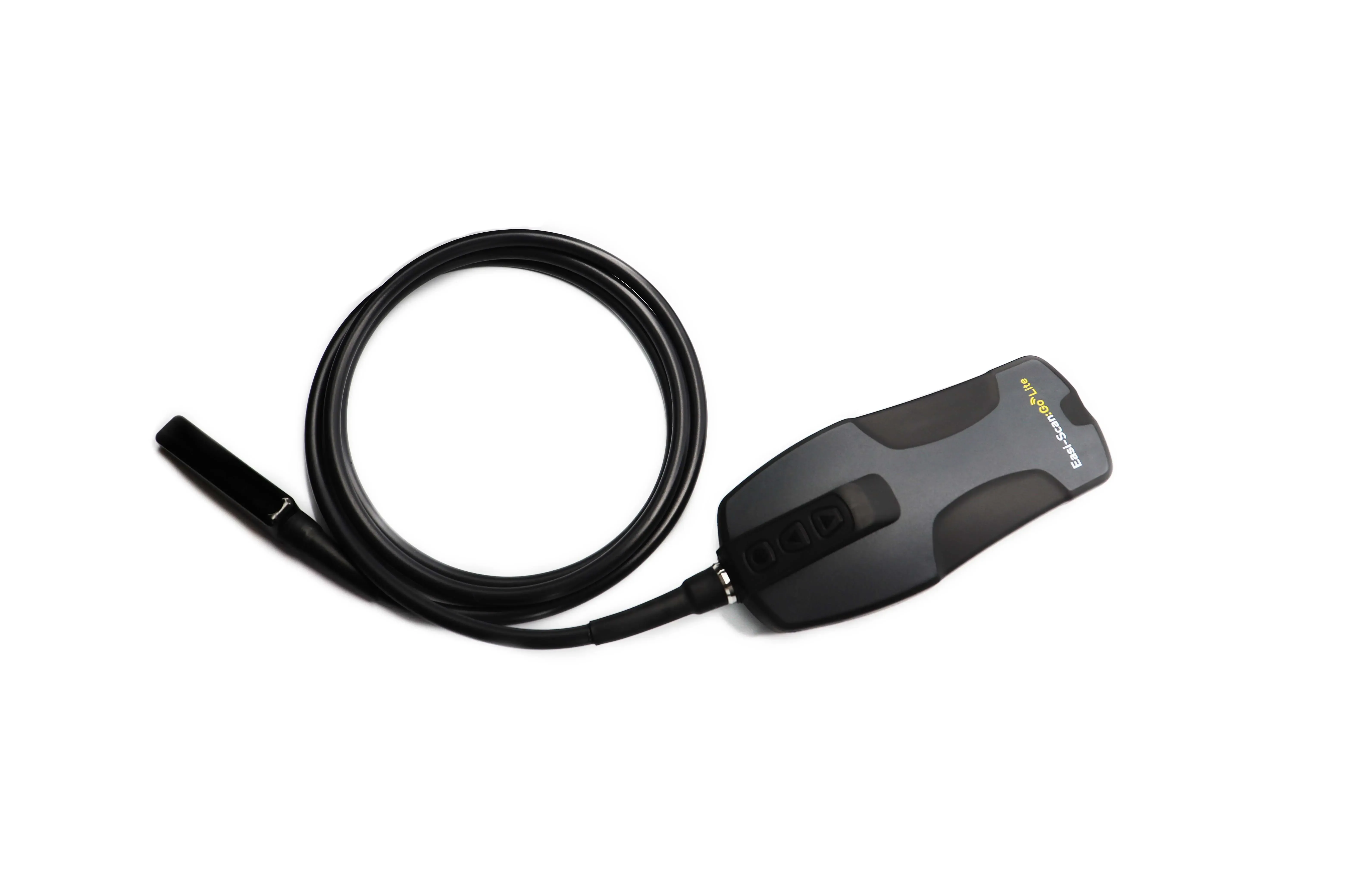
04.02.2022
Although Ultrasound has been used in cattle since 1980’s,it was still a new technique for routine ultrasound evaluation in the mid-90s. Moreover, at that time ultrasound devices were very heavy, lacked good image quality and came with very little available training.
Also at that time, some veterinarians and AI technicians started to use ultrasound in addition to rectal hand palpation and very quickly those service providers, as well as the dairy and beef producers understood the benefits of ultrasound – “seeing is believing” , the title name of DVM Paul R. Biagiotti article on the topic.
Practical applications of ultrasound are very wide ranging and include early assessment of pregnancy status, identification of cows carrying twins, detection of ovarian and uterine pathologies, and determination of fetal sex. Education programs to train bovine practitioners to use ultrasound for routine reproductive examinations are more and more available and definitively help them to provide a reliable diagnosis and treatment plans.
Ovarian Follicles : Most veterinary grade ultrasound scanners can identify ovarian follicles with a diameter of 2 to 3 mm or greater, and larger antral follicles (capable of ovulation) can easily be tracked during routine scanning sessions.
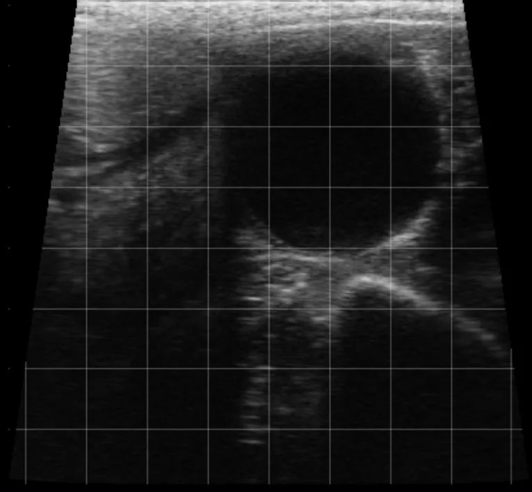
Image of an ovarian follicle taken with Easi-Scan:Go
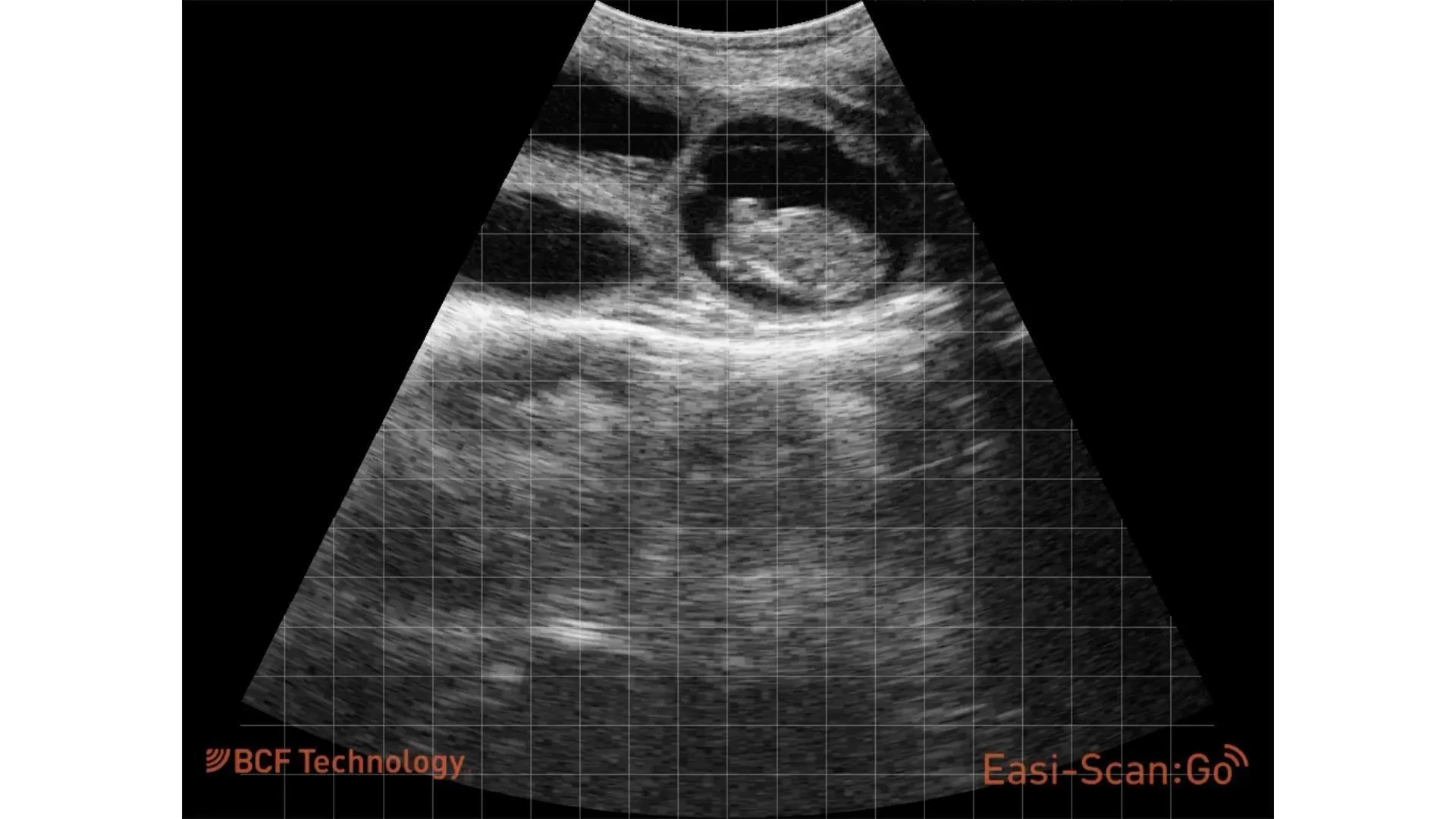
Corpus Luteum : Forms after ovulation from the tissues that constituted the ovarian follicle. Thus, the CL can be considered as the last stage of the follicular development. Several CL appear as solid tissue masses but may also contain fluid-filled cavities (79% in dairy heifers) from less than 2 to greater than 10 mm in diameter.
Ultrasound is a highly accurate and rapid method for assessing ovarian structures. The presence or absence of a corpus luteum aids in diagnosing pregnancy status. When present, the size and location (left or right) of the CL indicate the location of the conceptus within the uterus if the cow is pregnant. The presence of multiple CL is a great indicator for cows at risk for conceiving twins.
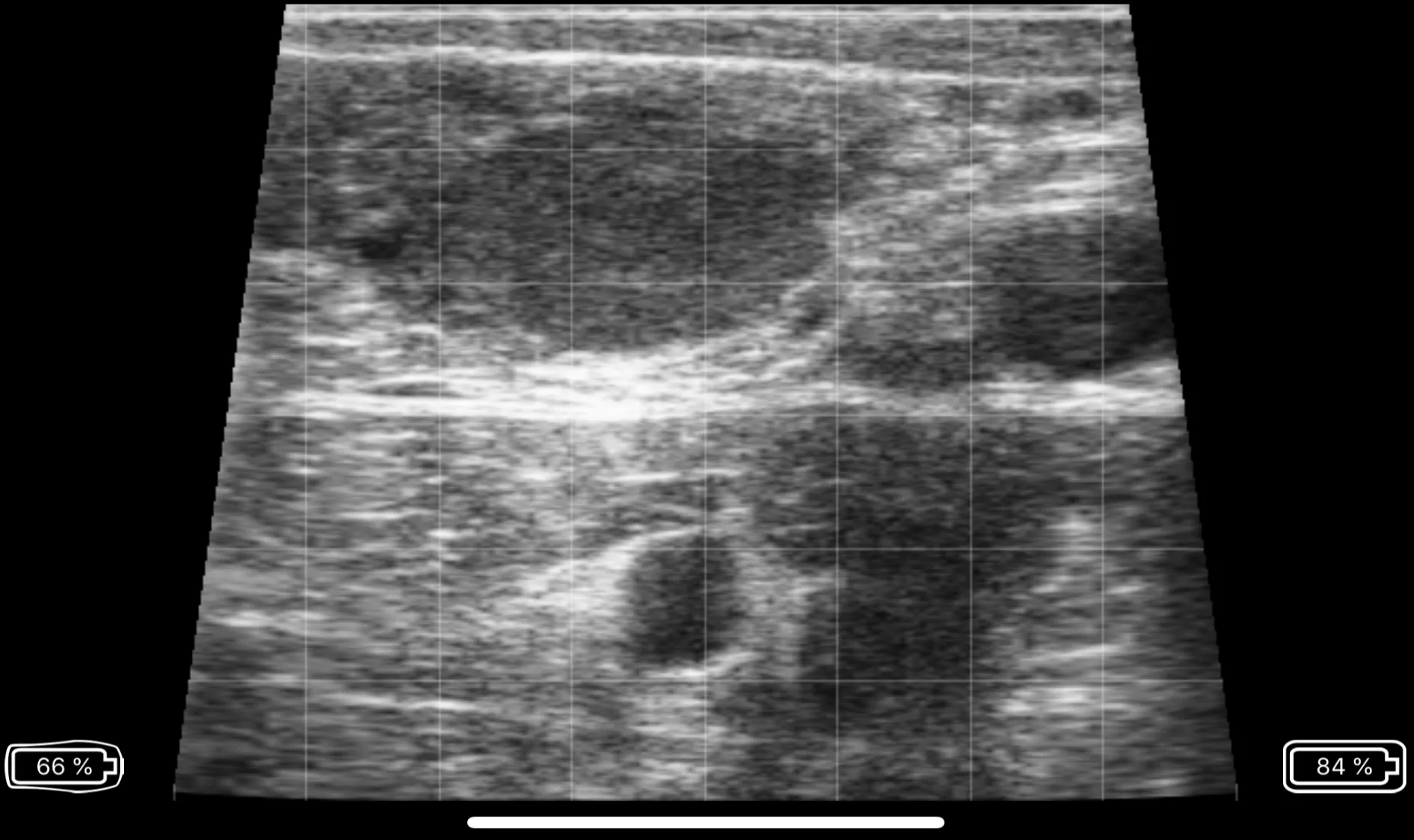
Image of corpus luteum taken with Easi-Scan:Go
Ovarian Cysts: Differentiation between follicular and luteal cysts via rectal palpation is difficult. When using transrectal ultrasonography, accuracy of the diagnosis is about 90% for luteal and nearly 75% for follicular cysts.
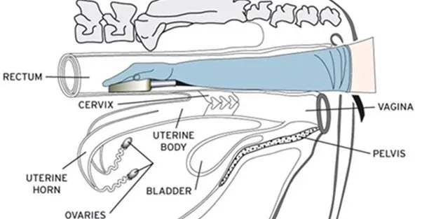
Scanning the Uterus
Early Pregnancy Diagnosis : One of the main application of ultrasound for cattle reproduction. Early identification of non-pregnant cows post-breeding improves reproductive efficiency and pregnancy rate. Under most on-farm conditions, pregnancy diagnosis can be accurately diagnosed using ultrasound as early as 26 days post AI, care must be taken to ensure the accuracy of a diagnosis. Sensitivity and specificity of pregnancy diagnosis are higher when ultrasound is conducted between 26 and 33 days post AI.
65 day pregnancy taken with Easi-Scan:Go Curve
In addition to early pregnancy diagnosis, fetal heart beat can be detected from 26 days and as gestation progresses, as the fetal thorax can be visualized, fetal heart beat and fetal viability can be also assessed.
Use of Colour Doppler:
Today, Doppler can be used for the diagnosis of non-gestation. Doppler can also be used to differentiate corpus luteum, luteinised follicular structure (very useful in case of super ovulated cows) and also to select recipients for embryo transfer.
Early Embryonic Loss: Of cows diagnosed pregnant at 28 days after AI, around 15-16% experience early embryonic loss by 56 days. Therefore, cows diagnosed pregnant at 28 days after AI should be scheduled for another examination around 60 days post AI, when the rate of embryonic loss per day decreases dramatically; it is also a good stage to do fetal sexing. Ultrasound is less invasive than rectal palpation and minimises the incidence of palpation-induced abortions.
Twin diagnosis: Cows carrying twin pregnancies can be accurately identified using transrectal ultrasonography 40 to 55 days after AI; the entire length of both uterine horns must be carefully scanned to ensure that an embryo is not missed. Because the majority of twins in dairy cattle occurs due to double ovulations, the presence of two or more CL on the ovaries is an excellent indicator of cows with an increased risk of conceiving twins. Several management scenarios could be considered upon identification of a cow carrying twins including culling, abortion and rebreeding, or continued management until parturition.
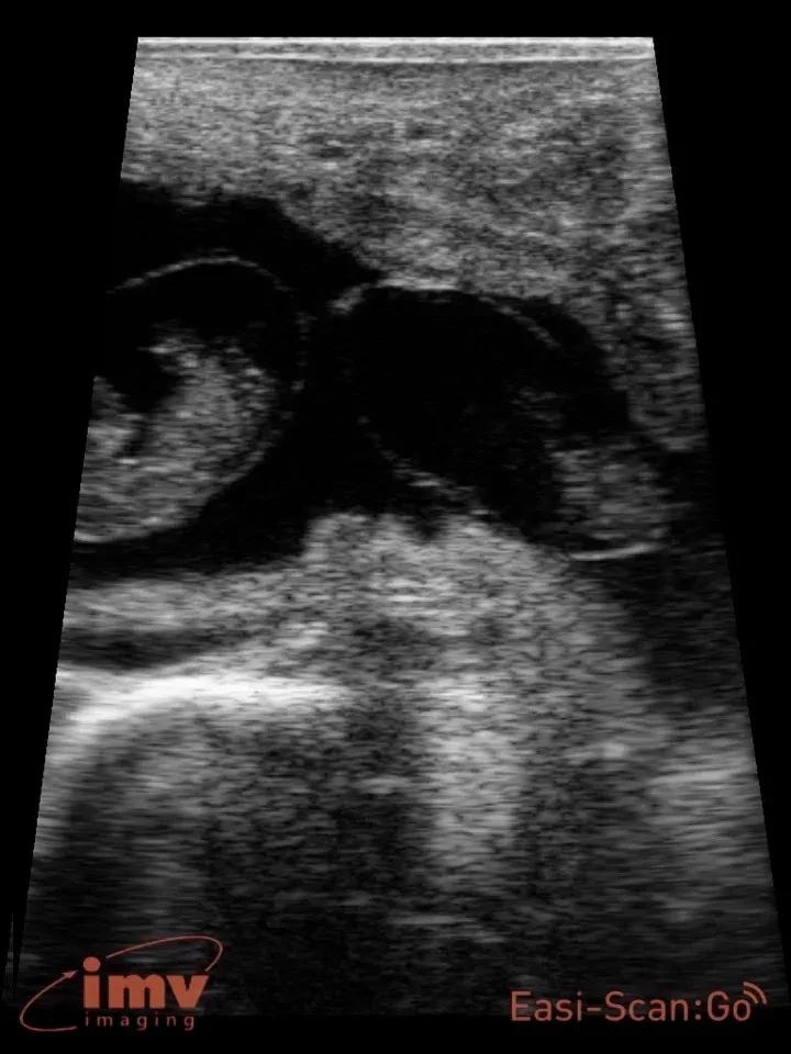
Image of Twins taken with Easi-Scan:Go
Fetal Sex: Sex is determined by evaluating the morphology and location of the genital tubercle and is a reliable and accurate method for sex determination beginning on days 55 to 60 of gestation.
{% video_player “embed_player” overrideable=False, type=’scriptV4′, hide_playlist=True, viral_sharing=False, embed_button=False, autoplay=False, hidden_controls=False, loop=False, muted=False, full_width=False, width=’464′, height=’640′, player_id=’65430567551′, style=” %}
60 day Male fetus taken with Easi-Scan:Go Curve
Calf’s Lungs scanning: it is a fast, non-invasive exam that provides a wealth of information quickly. You can see healthy lungs, pleural effusion, atelectasis, consolidation and abscessation. In a healthy lung, it is full of air and the lung does its job. In a diseased one, the surface of the lung is abnormal and thus there is penetration of the ultrasound waves deep into the lung parenchyma.
Udder and teat scanning: ultrasound can be used as a helpful tool to diagnose pathological alterations in the udder such as inflammation, mucosal lesions, tissue proliferation, foreign bodies, milk stones, congenital changes, hematoma and abscesses. Udder and teat scanning can be also performed for diagnosis of milk flow disturbances and different inner anatomical structures of the teat like teat canal length and diameter, teat cistern diameter and teat wall thickness.
Estimation of Fetal Age: Can be accomplished with good accuracy. The earlier in gestation that fetal ageing is performed, the more accurate the predicted calving date. Crown to rump length and head or trunk diameter are possible methods to age the fetus. Depending on the experience of the practitioner, the diagnosis could be very accurate up to 90-100 days. This application is very popular in Beef Cattle as most of females are mated naturally with bulls.
In addition research and commercial applications for ultrasound guided OPU and IVF are becoming more readily available to beef and dairy producers, worldwide and is now widely offered on-site. Oocytes are collected from donors on the farm of origin and the oocytes are transported to the IVF lab, cultured, and fertilized. After sufficient development of the resulting IVF embryos, the embryos are then shipped back to the farm of origin, to be transferred into recipient females. This significantly increases the opportunities of OPU/IVF for many clients, while keeping expenses relatively reasonable.
Another commercial application of OPU/IVF is the production of embryos for transfer to a problem breeder or stressed lactating dairy cows.
Ultrasound-guided OPU can be performed in pregnant cows, to a maximum of 100 days of gestation. Thus, a pregnant donor cow can still produce embryos, over a greater proportion of her lifetime.
This technology can be a valuable tool for seedstock producers interested in improving the carcass merit of their cattle. Carcass traits are moderately to highly heritable.
The measurements collected through live animal carcass ultrasound can be used to estimate carcass retail yield and meat quality. The common traits estimated include ribeye area (REA), rib fat (BF), rump fat (RF) and percent intramuscular fat (IMF%). Ribeye area is measured between the 12th and 13th ribs and gives an estimate of the amount of muscle and lean product in the animal. Rib fat (back fat) is also measured between the 12th and 13th ribs and is an estimate of the external fat on the animal. Rump fat is an additional measure of external fat on the animal. Marbling, or intramuscular fat, is the estimate of the amount of fat present in the loin / ribeye (longissimus dorsi) muscle. Graders evaluate the amount of intramuscular fat at the cut surface of the ribeye on the 12th rib surface.
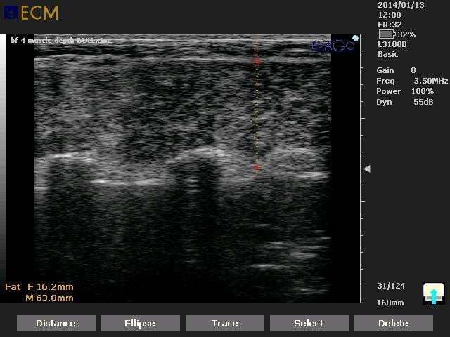
Back Fat and Muscle on bovine taken with ExaGo
Typical clinical ultrasonographic examinations of bull testes are unlikely to affect semen quality or sperm production. The ultrasonographic anatomy of bull testes and accessory sex glands is of great interest. A thorough history, complete physical examination, and a BSE (Breeding Soundness Evaluation) should precede an ultrasound examination of the bull’s genital system.
The scrotum and testes are then scanned in both a sagittal and transverse plane with the probe applied to the cranial, lateral, or caudal surface of the testis depending on the examiner’s preference. A complete examination includes the spermatic cord, the entirety of the testicular parenchyma, the epididymis, and the scrotal wall.
The testis parenchyma appears as a homogeneous tissue with a stippled medium echogenicity that surrounds the centrally located and more hyperechoic (whiter) mediastinum testis. The mediastinum testis is normally less than 5 mm wide. Changes in the ultrasonic appearance of the testis parenchyma can reflect normal events around puberty as well as pathology.