06.08.2024
Ultrasonography of the thorax may enable the pleural surface of the lung and superficial lung parenchyma, heart and mediastinal region to be evaluated.
Ultrasonography of the thorax may enable the pleural surface of the lung and superficial lung parenchyma, heart and mediastinal region to be evaluated. This is dependent on the species being examined, animal size and approach utilized.
How to scan
When evaluating the lung and pleural surface. The probe is initially placed in the intercostal space immediately caudal to the point of the elbow. And moved along the intercostal space in the dorsoventral direction (parallel to the ribs). See Figures 1 and 2 for probe placement in cattle and sheep.
Evaluation of the heart and pericardial space may be achieved in some animals. Particularly cattle which are in a thin body condition and sheep.
Pulling the leg forward can be helpful to reposition the location of the triceps muscle mass. Allowing the probe to be placed further ventrally and cranially to overly the cardiac region. The mediastinal space may also be visualized by placing the probe in the region of the thoracic inlet and directed causally.
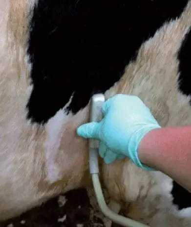
1 – Bovine thorax probe placement
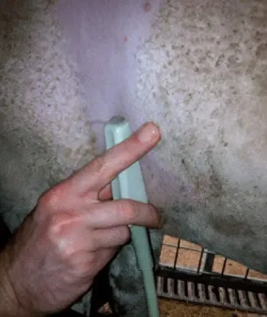
2 – Ovine thorax probe placement
Normal description
The presence of air within the normal lung prevents the ultrasound waves from penetrating deeply into the lung parenchyma. Therefore, the highly reflective pleural surface which is in contact with aerated lung. Is readily identified on ultrasound as a uniformly hyperechoic line which moves in synch with respiration. Figure 3 shows the surface of the lung in a normal cow with no evidence of pulmonary disease.
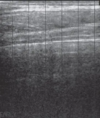
3 – Normal ultrasound appearance of the bovine thorax
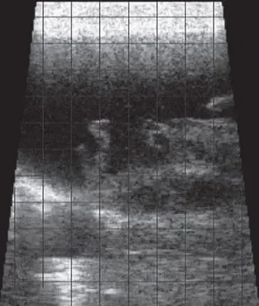
4 – Sheep with fibrinous pleuritis
Pathology
Conditions such as bovine respiratory disease (BRD)/chronic suppurative pulmonary disease (CSPD). Have a significant impact on health and productivity of both beef and dairy cattle. Ultrasound may be utilized in conjunction with clinical examination findings. Including demeanor, feed intake, rectal temperature, thoracic auscultation, and presence of coughing and nasal discharge to further evaluate the lungs for presence and distribution of lesions.
Figure 4 shows a sheep with fibrinous pleuritis. This is visualized as the presence of hypoechoic fluid within the thoracic cavity containing strands/aggregates of more hyperechoic material.
Why ultrasound?
Ultrasound may be used to identify abnormalities. Such as pleural effusion, septic pericarditis, lung lobe consolidation, ovine pulmonary adenocarcinoma and fibrinous pleuritis, as previously demonstrated.