28.01.2021
Ultrasound technology has been used in the reproductive management of sheep and goats since the 1960s.
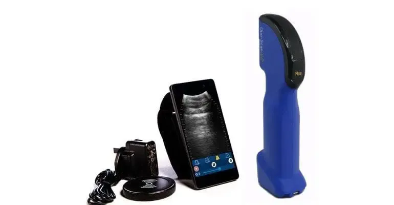
Recent advances in B-mode units in terms of portability, ease of contact lubrication application*, and more recently wireless streaming of ultrasound images on tablets and phones, have increased the speed and convenience of scanning.
Probe Positioning
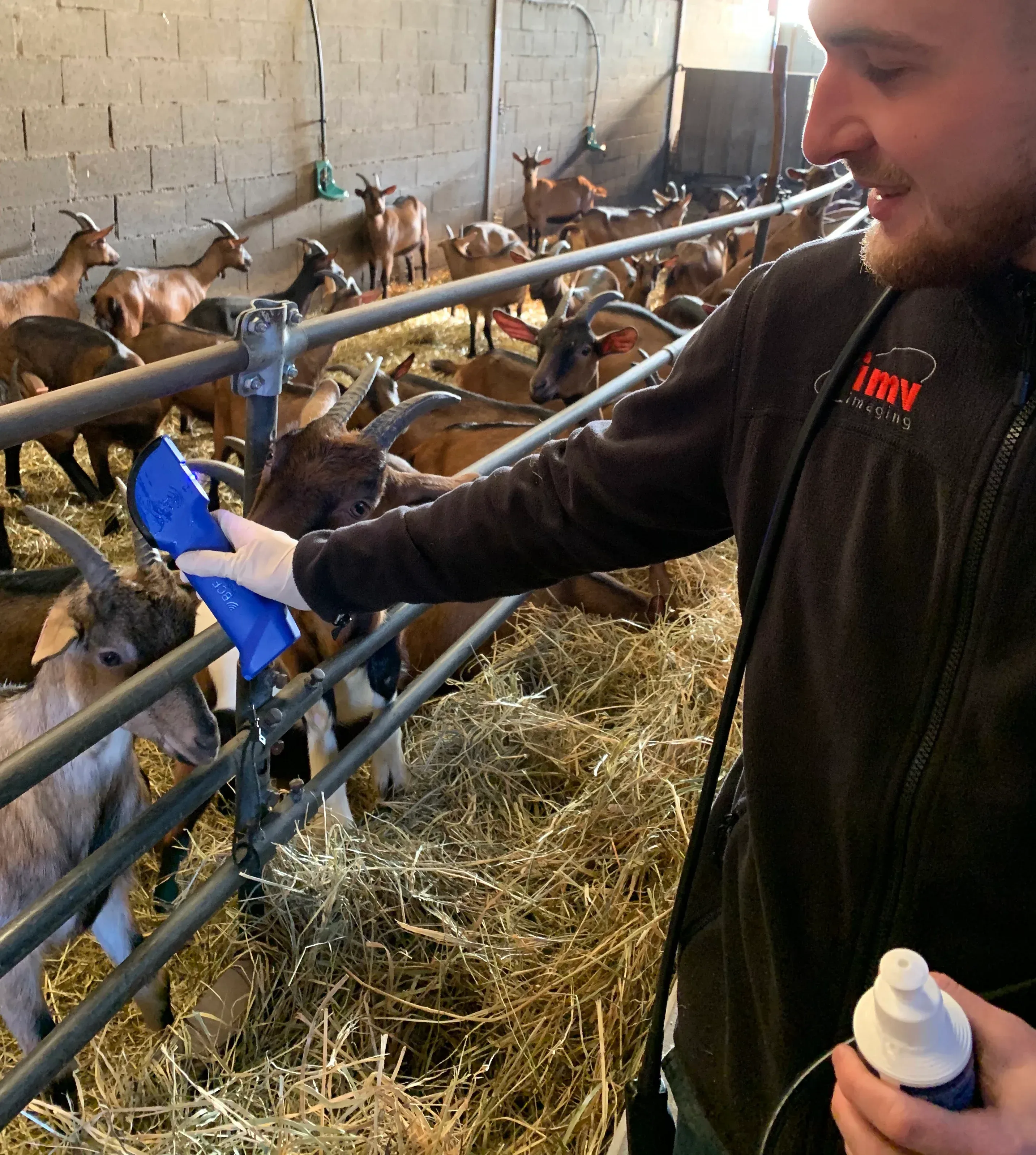
Probe placement worked best under the back leg, left or right, pointing back towards the midline of the doe and towards bladder. As with all animal scanning, the first point is achieving good contact between the transducer and the skin. If there are large black areas vertically down your image, usually at the sides, this means you are not making enough contact with the skin. The entire sector of the probe should be used. Very slight movements are required to bring the fetus into view. Practice keeping the head of the transducer in more or less the same spot and angling your probe back and forth using your wrist, as opposed to moving your whole arm around.
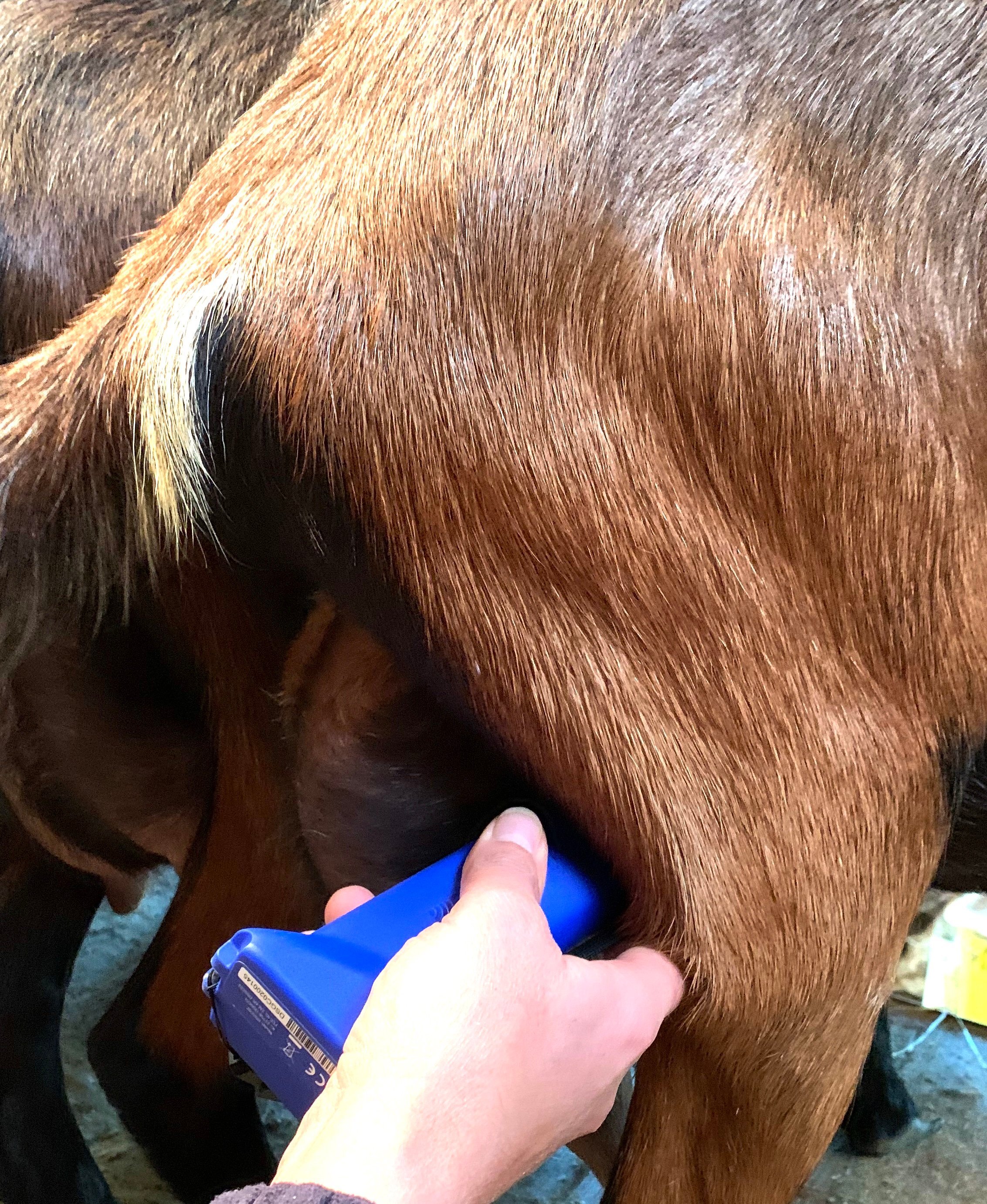
Transabdominal ultrasound scanning of uterus preferably takes place with the goats in a standing position. It is best to perform the examinations while the goats are standing in the milking stable or on the milking parlour because one can easily apply the transducer in the standing position and does not need to bend down. In addition, several goats can be tested close to each other and can be given some food so that they stand peacefully.
Applications
For pregnancy detection, transabdominal scanning of goats allows for a very efficient and accurate diagnosis. Experienced scanners’ users with highly efficient handling systems can scan as many as about 250 animals per hour at a 98% accuracy rate. The ideal window of pregnancy for transabdominal scanning is day 40-70.
{% video_player “embed_player” overrideable=False, type=’scriptV4′, hide_playlist=True, viral_sharing=False, embed_button=False, autoplay=False, hidden_controls=False, loop=False, muted=False, full_width=False, width=’360′, height=’640′, player_id=’41058573378′, style=” %}
Video shows a 55 day goat fetus with the Duo-Scan:Go Plus.
Producers interested in this application are advised to limit buck exposure to no more than 2 estrus cycles (42 days in goats) if a single scanning session is desired. Ideally, one would schedule scanning 40 days following buck removal.
Stage of pregnancy can be quantified with accuracy (within ±4 days of gestational age for an experienced practitioner during mid-pregnancy) with transabdominal scanning. Highly experienced practitioners are able to look at the relative proportions of placentome size (component of the goat placenta) to fluid space volume to fetal size along with changes in density of fetal bones to predict stage of pregnancy with a high degree of accuracy.
Fetal number can be quantified with reasonable accuracy in the day 40-70 stage of pregnancy as well. Skilled practitioners have error rates less than 5% in prediction of fetal number, although accuracy is hard to quantify from birth records due the natural occurrence of fetal death and reabsorption following the time of scanning in mid-pregnancy to term. Prediction of fetal number in higher order multiples (triplet or greater pregnancies) also has a higher degree of error compared to single or twin pregnancies. Accurate quantification of fetal number also takes more time than simple yes/no pregnancy detection, slowing animals scanned per hour to <50.
Pathologies
Hydrometra
Hydrometra is a pathological condition of the uterus in which accumulation of aseptic fluid in the uterine lumen takes place in the presence of a persistent corpus luteum. Pseudopregnancy is regarded as being synonymous; cases of hydrometra are found in mated or inseminated goats which remain anoestrous afterwards and are therefore thought to be pregnant. The characteristic ultrasonographic image of a hydrometra shows non-echogenic fluid compartments separated by double-layered thin tissue walls.
Fetal Death
The absence of fetal movements and heartbeats are the first ultrasonographic signs of fetal death. If degeneration of the fetus occurs, the ultrasound image shows an undifferentiated image of the uterine content with anechoic to hyperechoic structures. It is remarkable that the fetal membrane stays intact much longer than the fetus(es). Finally, this process can result in an ultrasonographic image that is identical to that of pyometra. If fetal death occurs at a more advanced stage of pregnancy, mummified fetuses or remnants of fetal bones show echogenic properties consistent with high tissue density.
Endometritis/Pyometra
In the goat, accumulation of cloudy fluid in the uterus can be associated with pathological conditions such as fetal death during early pregnancy or post-partum pyometra, however it is very rare.
Benefits
Better allocation of feed resources
Separation of ewes or does according to pregnancy status allows removal of non-pregnant animals, which then can either be culled or fed less due to their lower requirements. In more prolific flocks/herds, grouping according to fetal number results in a better allocation of feed resources, particularly during late pregnancy. Those carrying singles can be fed less, with more and higher quality feed resources reallocated to those rearing multiples. This will result in a better pregnancy outcome for the entire flock/herd.
Precision feeding to meet animal requirements during pregnancy
Animals can be fed precisely according to stage of pregnancy and litter size. Grouping animals using this information and feeding accordingly will reduce the incidence of metabolic disease and optimize maternal and fetal nutrition.
Optimizing birth management
Grouping according to fetal number and stage of pregnancy provides an opportunity to increase labor efficiency during the birth period. By separating does with potentially greater needs for labor at birth (those with multiples) from those with less (those carrying singles), one can allocate resources appropriately to achieve best birth outcomes.
Decision as to when to dry-off lactating animals in parlor milk production
The decision to dry-off lactating animals in parlour milk production can be made with knowledge of pregnancy status and stage. This allows animals to be managed in such a way as to allow optimal dry period length for better lactational performance and herd/flock health.
Detection of pregnancy in primiparous animals
Early detection of pregnancy allows sale of non-pregnant animals when they are still at a high market value (under 12 months of age). The ability to breed before one year of age can be used as a selection tool for increased lifetime productivity, as it has been demonstrated that does that kid at 12-15 months of age have a higher lifetime productivity than those that do not.
*Portable improvements in terms of battery powered options with head-mounted viewing displays. Lubrication contact advancements are down to some units allowing a stream of water or gel to be pumped over the probe.